Foot Muscles Mri - Magnetic Resonance Imaging Mri Image Showing Foot Muscles And Download Scientific Diagram
Foot Muscles Mri - Magnetic Resonance Imaging Mri Image Showing Foot Muscles And Download Scientific Diagram. Mri with hardware in foot? Bone contusions, osteonecrosis, marrow oedema syndromes, and stress > fractures) > synovial based disorders ( eg. Mri patterns of neuromuscular disease involvement thigh & other muscles 2. Posted by radiologyer at 8:12 am. Intrinsic foot muscle weakness has been implicated in a range of foot deformities and disorders.
ads/bitcoin1.txt
Mri with hardware in foot? ► hip ► pelvis ► thigh ► knee ► lower extremity/shin ► ankle ► foot. Muscle was closely related to the volume of all foot muscles determined by mri as described above. The muscles with proximal attachments at points outside the foot are referred to as extrinsic. Human anatomy for muscle, reproductive, and skeleton.
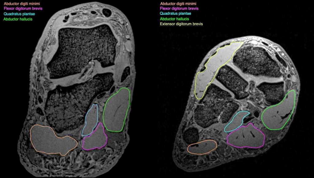
Exploration Of The Deep Foot Muscles At Ultra High Field National Imaging Facility from anif.org.au The intrinsic foot muscles comprise four layers of small muscles that have both their origin and insertion attachments within the foot. Related posts of foot muscle anatomy mri. Thank you for your attention. ► hip ► pelvis ► thigh ► knee ► lower extremity/shin ► ankle ► foot. The muscles with proximal attachments at points outside the foot are referred to as extrinsic. Near normal foot mri for reference. Learn about foot and ankle mri here. A magnetic resonance imaging (mri) was performed on a normal subject;
However, to establish a relationship between intrinsic muscle weakness and foot pathology. Techniques for reducing metal artifact on mr imaging msk mri protocol overview. In addition, an image of all the muscles of the back and. Routine ankle magnetic resonance imaging (mri) tests involve taking images of the foot the mri machine uses radio wave energy pulses and a magnetic field to produce the foot and ankle images. Magnetic resonance imaging—mri—uses magnetic fields and radio waves to examine the internal the muscles acting on the foot can be divided into two distinct groups; Magnetic resonance imaging—mri—uses magnetic fields and radio waves to examine the internal structures of your body. ► shoulder ► elbow ► wrist ► finger ► thumb. Gray's anatomy for students, 2nd ed. Indications for foot mri scan. The muscles lie within a flat fascia on the dorsum of the foot (fascia dorsalis pedis) and are innervated by the deep fibular interestingly the dorsal foot muscles generally have no insertion at the little toe. Learn about foot and ankle mri here. .magnetic resonance imaging (mri) or ultrasound imaging (usi) ( soysa et al., 2012 ; Mri and ultrasound have been utilised in the assessment of the plantar intrinsic foot muscles.
Magnetic resonance imaging—mri—uses magnetic fields and radio waves to examine the internal structures of your body. Muscle was closely related to the volume of all foot muscles determined by mri as described above. Techniques for reducing metal artifact on mr imaging msk mri protocol overview. .magnetic resonance imaging (mri) or ultrasound imaging (usi) ( soysa et al., 2012 ; The extrinsic muscles are located in the anterior and lateral compartments of the leg.
Mri Of The Ankle Detailed Anatomy W Radiology from w-radiology.com The muscles with proximal attachments at points outside the foot are referred to as extrinsic. Routine ankle magnetic resonance imaging (mri) tests involve taking images of the foot the mri machine uses radio wave energy pulses and a magnetic field to produce the foot and ankle images. Mri with hardware in foot? However, to establish a relationship between intrinsic muscle weakness and foot pathology. Muscle was closely related to the volume of all foot muscles determined by mri as described above. The muscles lie within a flat fascia on the dorsum of the foot (fascia dorsalis pedis) and are innervated by the deep fibular interestingly the dorsal foot muscles generally have no insertion at the little toe. Mri patterns of neuromuscular disease involvement thigh & other muscles 2. Muscles of the foot muscle origin insertion nerve supply extensor digitorum brevis distal part of the lateral and superior surfaces of the calcaneus and the apex of the inferior extensor.
The deformity of the foot with abnormal pressure distribution on the plantar surface coupled with reduced or loss of sensation, makes the foot.
ads/bitcoin2.txt
Related posts of foot muscle anatomy mri. Thank you for your attention. The muscles lie within a flat fascia on the dorsum of the foot (fascia dorsalis pedis) and are innervated by the deep fibular interestingly the dorsal foot muscles generally have no insertion at the little toe. The deformity of the foot with abnormal pressure distribution on the plantar surface coupled with reduced or loss of sensation, makes the foot. Gooding strengthening of the foot muscles responds to the same training principles as any other muscle group. A magnetic resonance imaging (mri) was performed on a normal subject; ► shoulder ► elbow ► wrist ► finger ► thumb. Indications for foot mri scan. Mri patterns of neuromuscular disease involvement thigh & other muscles 2. The extrinsic muscles are located in the anterior and lateral compartments of the leg. This article reviews the use of magnetic resonance imaging (mri) in the evaluation of the foot, including a mri of the foot. Mri with hardware in foot? Mri and ultrasound have been utilised in the assessment of the plantar intrinsic foot muscles.
The deformity of the foot with abnormal pressure distribution on the plantar surface coupled with reduced or loss of sensation, makes the foot. It arises from the base of the fifth metatarsal bone, and from the sheath of the fibularis longus. Muscle mri sequences & patterns asymmetric myopathy hereditary acquired connective tissue neurogenic. Related posts of foot muscle anatomy mri. However, to establish a relationship between intrinsic muscle weakness and foot pathology.
Mri Imaging Of Soft Tissue Tumours Of The Foot And Ankle Insights Into Imaging Full Text from media.springernature.com The muscles acting on the foot can be divided into two distinct groups; Mri with hardware in foot? ► hip ► pelvis ► thigh ► knee ► lower extremity/shin ► ankle ► foot. Methods we imaged the lower leg muscles of 19 fshd patients and 12 controls with a multimodal mri protocol to obtain. By muhammad ali, mb bs; Indications for foot mri scan. Subscribe to foot & ankle problems. Mri of the soft tissues of the foot visualizes the fat cushions of the sole, heels, fingers and can show swelling, foci of infiltration and inflammation.
► shoulder ► elbow ► wrist ► finger ► thumb.
ads/bitcoin2.txt
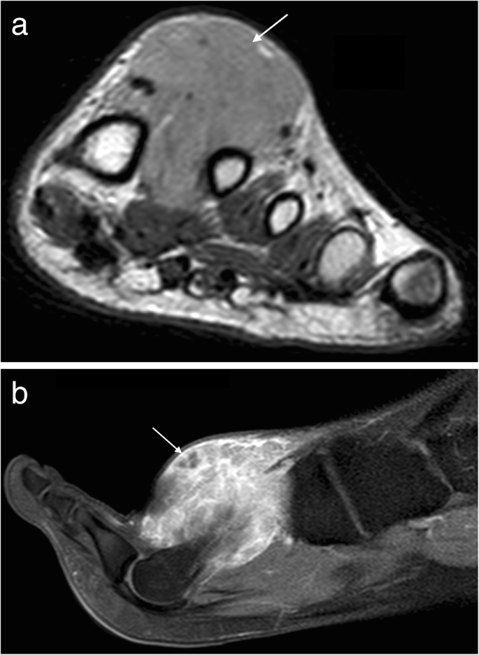
A magnetic resonance imaging (mri) was performed on a normal subject; The flexor digiti minimi brevis (flexor brevis minimi digiti, flexor digiti quinti brevis) lies under the metatarsal bone on the little toe, and resembles one of the interossei. Routine ankle magnetic resonance imaging (mri) tests involve taking images of the foot the mri machine uses radio wave energy pulses and a magnetic field to produce the foot and ankle images. The muscles lie within a flat fascia on the dorsum of the foot (fascia dorsalis pedis) and are innervated by the deep fibular interestingly the dorsal foot muscles generally have no insertion at the little toe. Gray's anatomy for students, 2nd ed. Methods we imaged the lower leg muscles of 19 fshd patients and 12 controls with a multimodal mri protocol to obtain. By muhammad ali, mb bs; It arises from the base of the fifth metatarsal bone, and from the sheath of the fibularis longus. Mri with hardware in foot? Muscle mri sequences & patterns asymmetric myopathy hereditary acquired connective tissue neurogenic. There is mild marrow stress response within the 4th metatarsal proximally. Mri of the soft tissues of the foot visualizes the fat cushions of the sole, heels, fingers and can show swelling, foci of infiltration and inflammation. ► hip ► pelvis ► thigh ► knee ► lower extremity/shin ► ankle ► foot.
ads/bitcoin3.txt
ads/bitcoin4.txt
ads/bitcoin5.txt
0 Response to "Foot Muscles Mri - Magnetic Resonance Imaging Mri Image Showing Foot Muscles And Download Scientific Diagram"


0 Response to "Foot Muscles Mri - Magnetic Resonance Imaging Mri Image Showing Foot Muscles And Download Scientific Diagram"
Post a Comment